
Rapid Alteration of Buccal Cortical Bone following Acute Apical Abscess: Implications for Endodontic Microsurgery: A Case Report
Received 27 May 2026
Revised following the first round of review 25 September 2026
Revised following the second round of review 16 October 2026
Accepted following the third round of review 25 October 2026
Published online 31 October 2026
J Endod Microsurg. 2026;5: 100021.
Under a Creative Commons license
HOW TO CITE THIS ARTICLE: AMA STYLE
Shahravan A, Moradinia M, Jamshidi Jam N, Abbasi Sardari A. Rapid alteration of buccal cortical bone following acute apical abscess: Implications for endodontic microsurgery: A case report. J Endod Microsurg. 2026;5:100021.

ABSTRACT
Introduction: In endodontic microsurgery, cone-beam computed tomography (CBCT) imaging serves as a roadmap for assessing buccal cortical bone thickness and planning the osteotomy. However, periapical lesions are dynamic in nature. This case report describes a discrepancy between preoperative CBCT findings, and the surgical anatomy observed after an acute apical abscess.
Case Report: A 25-year-old female presented with persistent pain and swelling in the area of tooth #19 (mandibular first molar). Initial CBCT (T₀) showed a relatively thick buccal cortical plate overlying the periapical lesion, with no apparent cortical perforation in the available images. After an acute flare-up with severe swelling just before the scheduled surgery, intraoperative observation revealed extensive buccal cortical fenestration. The surgery was completed under microscopy, including retrieval of a separated file, isthmus preparation, and retrograde root-end filling with a bioceramic material (NeoPutty, Avalon Biomed, Houston, TX, USA). At the one-year follow-up, the periapical radiograph demonstrated substantial radiographic healing.
Conclusion: This case highlights a discrepancy between preoperative CBCT findings, and the cortical anatomy encountered during surgery following an acute inflammatory exacerbation. Although the exact timing and mechanism of cortical destruction cannot be established from a single case without interval imaging, the findings emphasize that previously obtained CBCT images may not fully represent the surgical anatomy following an acute flare-up. Clinicians should therefore consider the possibility of altered cortical anatomy and be prepared to modify the surgical approach when acute clinical changes occur.
KEYWORDS
Acute apical abscess, endodontic microsurgery, buccal cortical bone, cone-beam computed tomography, surgical treatment planning.
INTRODUCTION
Among adults presenting with acute dental conditions, approximately 16% will have symptomatic apical periodontitis and a further 20% will have an acute apical abscess [1].
The integration of endodontic microsurgery (EMS) and cone-beam computed tomography (CBCT) has revolutionized the management of persistent periapical disease; EMS has demonstrated favorable clinical and radiographic outcomes when appropriate case selection and modern microsurgical techniques are employed. CBCT provides essential data regarding the three-dimensional extent of the lesion, its proximity to vital structures, and the thickness of the buccal cortical plate, which dictates the surgical approach [2].
However, clinicians must recognize that a periapical lesion is a dynamic biological entity rather than a static radiographic image. The transition from a chronic state to an acute exacerbation, described as an acute exacerbation of a pre-existing periapical lesion [3], involves a complex interplay of microbial aggression and host response. This phase is characterized by an influx of polymorphonuclear leukocytes (PMNs) and increased production of pro-inflammatory cytokines such as IL-1β, IL-6, and TNF-α, which can promote osteoclastic bone resorption [4].
The pattern and rate of bone destruction may differ between chronic and acute inflammatory lesions. Acute apical abscesses may be associated with accelerated inflammatory bone resorption [5]. In addition, accumulation of purulent exudate within confined tissue spaces may increase local tissue pressure and compromise microcirculation [6, 7]. Pressure-related ischemic injury is a proposed biological hypothesis, not an established explanation for cortical perforation in endodontic periapical disease; its contribution in the present case cannot be demonstrated.
Acute flare-ups may coincide with changes in local clinical conditions [8, 9]. Accordingly, the anatomy encountered during surgery may differ from that depicted on previously acquired imaging; this discrepancy alone does not establish when or why a cortical defect developed.
Therefore, in the present case, pressure-related ischemic injury is considered only as one possible mechanism rather than a definitive explanation for the observed cortical defect. The observed cortical defect may reflect several biological processes associated with inflammation, but neither its mechanism nor its timing can be established from the available evidence.
The present case report describes a patient in whom the available preoperative CBCT demonstrated an apparently intact buccal cortical plate, whereas extensive buccal cortical fenestration was identified intraoperatively following an acute clinical exacerbation. Because interval imaging was not available, the exact timing and mechanism of cortical destruction cannot be established. Nevertheless, the case illustrates a potential discrepancy between preoperative CBCT findings and intraoperative anatomy after an acute flare-up, without establishing that the flare-up caused the cortical defect.
CASE PRESENTATION
A 25-year-old female patient (ASA I, i.e., a normal healthy patient) presented with a complaint of constant pain and tenderness to percussion in tooth #19 (mandibular first molar). Her dental history revealed a root canal retreatment on that tooth 18 months prior. According to the previous records, a file fragment had separated in the apical part of the mesiolingual canal during the retreatment (Fig 1). Periodontal probing around tooth #19 revealed normal probing depths of approximately 1-2 mm, with no isolated deep periodontal pocket.


FIGURE 1. Preoperative intraoral photograph (A) and panoramic radiograph (B) showing the periapical lesion associated with the mesial root of tooth #19.
Initial Radiographic Evaluation: The initial CBCT imaging (T₀) confirmed the presence of a periapical radiolucent lesion around the mesial root of #19. The available preoperative CBCT images showed a relatively thick buccal cortical plate overlying the periapical lesion, with no apparent cortical perforation in the imaged sections (Fig 2). Table 1 an overview of the clinical stages, timing, and key findings/management.


FIGURE 2. Preoperative CBCT images: axial view at the middle third of the root (A), axial view at the apical third (B), axial view at the level of the root apex (C), coronal views of mesial root (D, E), coronal view of distal root (F), and sagittal views (G, H).
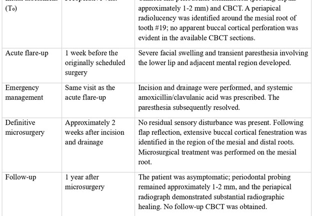
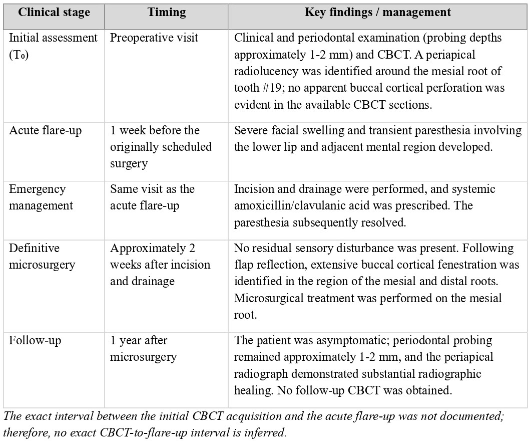
TABLE 1. Timeline.
Acute Flare-up: One week before the scheduled surgery, the patient returned with severe facial swelling and transient paresthesia involving the lower lip and adjacent mental region. Emergency treatment was performed, consisting of incision and drainage of the abscess and administration of systemic antibiotics (amoxicillin/clavulanic acid [Cosar Pharmaceutical Co., Tehran, Iran]). The paresthesia resolved following incision and drainage, and the patient reported no residual sensory disturbance at the time of the surgical appointment. The definitive microsurgery was postponed by two weeks to allow the acute infection and inflammation to resolve.
Surgical Procedure
The endodontic surgery was performed under local anesthesia and high-power magnification (operating microscope), with the following steps:
Anesthesia and Hemostasis: An inferior alveolar nerve block (2% Lidocaine with 1:80,000 epinephrine [Darupakhsh, Tehran, Iran]), complemented by buccal infiltration (4% Articaine with 1:200,000 epinephrine [Eksir, Tehran, Iran]), was administered to achieve profound anesthesia and hemostasis in the surgical area.
Flap Reflection: A full-thickness triangular flap with a mesial vertical releasing incision was designed. The flap was elevated using a Molt #9 periosteal elevator to expose the buccal surface of the bone.
Intraoperative Findings: Following flap reflection, an extensive buccal cortical defect was immediately identified in the region of both the mesial and distal roots of tooth #19 (Fig 3). The defect provided direct surgical access to the root apices, and therefore no conventional osteotomy was required. This finding differed substantially from the cortical anatomy demonstrated on the available preoperative CBCT images. Because no interval CBCT was obtained after the acute exacerbation, the exact timing of development of the cortical defect could not be determined.
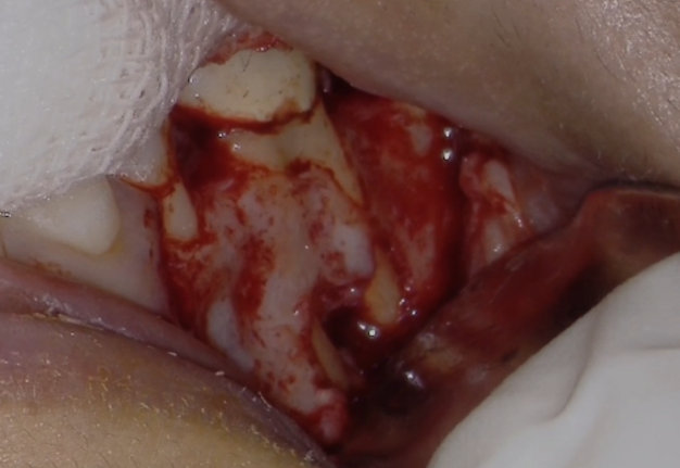
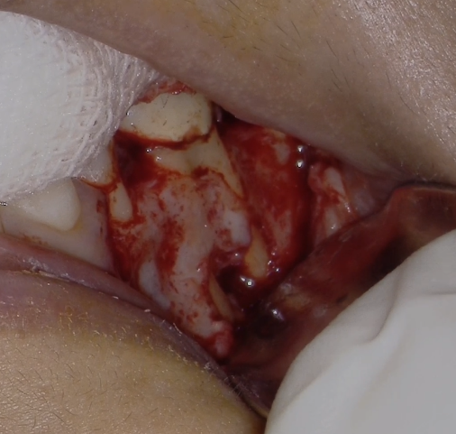
FIGURE 3. Intraoperative view demonstrating extensive buccal cortical fenestration in the region of the mesial and distal roots of tooth #19.
Biopsy and Curettage: All granulation tissue and associated bone fragments were thoroughly curetted. The tissue was fixed in 10% formalin and submitted for histopathological examination to rule out non-endodontic pathology and confirm the periapical inflammatory origin. Histopathological examination of the curetted periapical tissue demonstrated a periapical granuloma (Fig 4). No evidence of a non-endodontic pathological process was identified. This finding was consistent with the inflammatory periapical origin of the lesion observed clinically and radiographically.
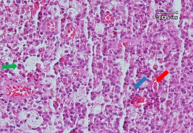
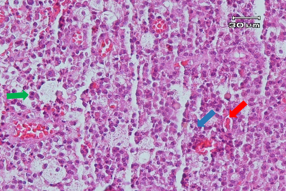
FIGURE 4. Histopathological section stained with hematoxylin and eosin (H&E), showing inflammatory cell infiltration (periapical granuloma). The blue arrow indicates a lymphocyte, the green arrow indicates a plasma cell, and the red arrow indicates erythrocytes. Scale bar = 30 µm.
Apical Management: The mesial root was resected by approximately 3 mm using a small round diamond bur. All granulomatous tissue in the periapical area was thoroughly removed with a curette. Although the cortical fenestration extended to the region of both roots, the distal root was not resected because no periapical lesion or other pathological finding requiring root-end surgery was identified at its apical region on the preoperative CBCT.
Removal of Separated File: The isthmus between the mesiobuccal and mesiolingual canals was refined using an ASLD ultrasonic tip (ACTEON®, Mérignac, France) under high magnification. The separated file fragment that was lodged in this isthmus was loosened with ultrasonic vibration and successfully retrieved (Fig 5).


FIGURE 5. Intraoperative view (A, B) following resection of the mesial root.
Retrograde Filling: Each root-end cavity was prepared to a depth of about 3 mm using an ASLD ultrasonic tip (ACTEON®, Mérignac, France). After achieving hemostasis with an epinephrine-soaked cotton pellet, a premixed bioceramic putty (NeoPutty, Avalon Biomed, Houston, TX, USA) was placed to seal the resected root-end cavities as well as the mesial root isthmus (Fig 6).


FIGURE 6. Root-end filling with premixed bioceramic putty (A, B).
Closure and Suturing: The flap was repositioned and sutured with 6–0 nylon (Supa Co, Tehran, Iran). Postoperative instructions were given, and the patient’s recovery was uneventful (Fig 7).
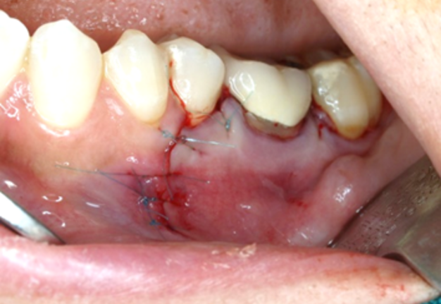
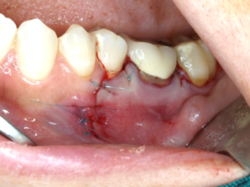
FIGURE 7. Flap repositioning and suturing with 6–0 nylon.
At the one-year follow-up (Fig 8), the patient was asymptomatic, and periodontal probing remained within normal limits (1-2 mm), with no isolated periodontal pocket associated with tooth #19. The periapical radiograph demonstrated substantial radiographic healing with re-establishment of the lamina dura around the root apices. Because no follow-up CBCT was obtained, complete three-dimensional regeneration of the buccal cortical plate could not be confirmed.


FIGURE 8. One-year follow-up periapical radiograph (A) and clinical photograph (B).
DISCUSSION
The key point in this case is the discrepancy between the buccal cortical anatomy demonstrated on the available preoperative CBCT and the extensive cortical fenestration identified intraoperatively following an acute flare-up. Although the initial CBCT demonstrated no apparent cortical perforation in the available sections and extensive cortical fenestration was identified intraoperatively after the acute flare-up, the absence of interval imaging prevents definitive determination of whether the entire cortical defect developed during this short interval. The temporal association between the acute exacerbation and the extensive cortical defect suggests a possible relationship; however, causality and the exact rate of cortical destruction cannot be established from a single case.
From a surgical perspective, the loss of the cortical plate provided direct access to the root apices, eliminating the need for a conventional osteotomy, but it also resulted in loss of bony support. Guided tissue regeneration (GTR) has been proposed as an adjunct to surgical endodontic treatment to enhance bone regeneration, particularly in cases involving large periapical lesions, through-and-through defects, or other challenging osseous defects. Previous systematic reviews and meta-analyses have suggested a potential benefit of GTR in selected clinical situations, although its benefit is not uniformly demonstrated across all cases [10]. In the present case, GTR was not considered necessary based on the clinical characteristics and morphology of the osseous defect. Nevertheless, regenerative procedures may be considered in selected cases depending on the extent, configuration, and morphology of the defect. The defect was managed with thorough curettage and primary flap closure.
A premixed bioceramic putty was used for root-end filling, and its handling characteristics allowed placement under the clinical conditions encountered. Premixed calcium silicate–based bioceramic putties have emerged as clinically applicable root-end filling materials in endodontic microsurgery. Previous studies have demonstrated favorable sealing properties of these materials when used for retrograde root-end filling [11]. Clinical evidence also supports their use; a retrospective CBCT-based cohort study reported high success rates following EMS using root repair material (RRM) putty, with no significant difference compared with MTA [12]. More recently, a randomized clinical trial demonstrated favorable periapical healing following the use of a premixed bioceramic putty as a root-end filling material [13]. Furthermore, experimental evidence suggests that premixed putty-type bioceramic materials can be effectively used alone for root-end filling [14]. Collectively, these findings support the clinical use of premixed bioceramic putties as an alternative root-end filling material in EMS.
An important limitation of this case is that the preoperative assessment of the buccal cortical plate was based on CBCT images obtained before the acute exacerbation. Although axial, coronal, and sagittal views are presented, interval CBCT imaging was not performed immediately before surgery; therefore, a pre-existing subtle cortical defect or interval progression cannot be completely excluded. Accordingly, although extensive cortical fenestration was observed intraoperatively following the acute flare-up, the exact timing and rate of cortical bone alteration cannot be established with certainty. In addition, the absence of standardized periodontal measurements at intermediate follow-up visits limits assessment of periodontal recovery over time.
The intraoperative finding of a large cortical defect also has practical implications for surgical planning. Loss of cortical support may alter flap management, access, hemostasis, and the potential need for regenerative procedures. In the present case, GTR was not considered necessary based on the clinical characteristics and morphology of the osseous defect. However, this approach may not be applicable to larger or anatomically different defects. The normal periodontal probing findings at one-year follow-up (1-2 mm) provided no clinical evidence of an isolated periodontal defect.
CONCLUSION
This case illustrates that the surgical anatomy of a periapical lesion may differ substantially from that represented on a previously acquired CBCT, particularly when a significant acute clinical exacerbation occurs before surgery. Although the exact timing and mechanism of the observed cortical defect cannot be established, clinicians should recognize that substantial anatomical changes may occur and should be prepared to reassess imaging and adapt the surgical approach when the clinical condition changes before endodontic microsurgery.
Author Contributions (CRediT)
Arash Shahravan: Conceptualization (lead); Investigation (lead); Writing – review & editing.
Mohammad Moradinia: Writing – original draft; Writing – review & editing.
Niloofar Jamshidi Jam: Writing – original draft; Writing – review & editing.
Ali Abbasi Sardari: Writing – original draft; Writing – review & editing.
AI Statement: ChatGPT (OpenAI) was used to assist with drafting and revising the manuscript, improving its English language and organization, and preparing responses to reviewers. The authors reviewed and edited the AI-assisted content and take full responsibility for the accuracy, integrity, and final content of the manuscript.
Declaration of Interests: The authors have no conflicts of interest to declare.
Informed Consent: Written consent was obtained from patient for publication of photographs.
Fundings: No funding was received for this study.
Peer-Review: Two external reviewers and one internal reviewer.
REFERENCES (14)
-
Cope AL, Francis NA, Wood F, Chestnutt IG. Antibiotic prescribing in UK general dental practice: a cross-sectional study. Community Dent Oral Epidemiol. 2016;44(2):145-153. https://doi.org/10.1111/cdoe.12199
-
Bieszczad D, Wichlinski J, Kaczmarzyk T. Treatment-related factors affecting the success of endodontic microsurgery and the influence of GTR on radiographic healing-a cone-beam computed tomography study. J Clin Med. 2023;12(19):6382. https://doi.org/10.3390/jcm12196382
-
Buonavoglia A, Latronico F, Pirani C, et al. Symptomatic and asymptomatic apical periodontitis associated with red complex bacteria: clinical and microbiological evaluation. Odontology. 2013;101(1):84-88. https://doi.org/10.1007/s10266-011-0053-y
-
Nair PN. Pathogenesis of apical periodontitis and the causes of endodontic failures. Crit Rev Oral Biol Med. 2004;15(6):348-381. https://doi.org/10.1177/154411130401500604
-
Jalali P, Tahmasbi M, Augsburger RA, et al. Dynamics of bone loss in cases with acute or chronic apical abscess. J Endod. 2019;45(9):1114-1118.
-
Langeland K. Tissue changes incident to cavity preparation. An evaluation of some dental engines. Acta Odontol Scand. 1961;19:397-430. https://doi.org/10.3109/00016356109043398
-
Schuknecht B, Valavanis A. Osteomyelitis of the mandible. Neuroimaging Clin N Am. 2003;13(3):605-618. https://doi.org/10.1016/s1052-5149(03)00044-3
-
Seltzer S, Bender IB. Cognitive dissonance in endodontics. Oral Surg Oral Med Oral Pathol. 1965;20(4):505-516. https://doi.org/10.1016/0030-4220(65)90244-6
-
Tsesis I, Faivishevsky V, Fuss Z, Zukerman O. Flare-ups after endodontic treatment: a meta-analysis of literature. J Endod. 2008;34(10):1177-1181. https://doi.org/10.1016/j.joen.2008.07.016
-
Tsesis I, Rosen E, Tamse A, et al. Effect of guided tissue regeneration on the outcome of surgical endodontic treatment: a systematic review and meta-analysis. J Endod. 2011;37(8):1039-1045. https://doi.org/10.1016/j.joen.2011.05.016
-
Rencher B, Chang AM, Fong H, et al. Comparison of the sealing ability of various bioceramic materials for endodontic surgery. Restor Dent Endod. 2021;46(3):e35. https://doi.org/10.5395/rde.2021.46.e35
-
Eskandar RF, Al-Habib MA, Barayan MA, Edrees HY. Outcomes of endodontic microsurgery using different calcium silicate-based retrograde filling materials: a cohort retrospective cone-beam computed tomographic analysis. BMC Oral Health. 2023;23(1):70. https://doi.org/10.1186/s12903-023-02782-w
-
Salah HM, Hashem AAR, Mustafa T, et al. The impact of root end filling material type and the application of bone graft on healing of periapical tissues after endodontic microsurgery (a clinical randomized controlled trial). Sci Rep. 2024;14(1):25378. https://doi.org/10.1038/s41598-024-66033-w
-
Knapp J, Kirkpatrick T, Ontiveros JC, et al. Efficacy of root-end filling techniques using premixed putty type bioceramic cements: an ex vivo study. Clin Oral Investig. 2024;28(7):365. https://doi.org/10.1007/s00784-024-05759-3