
Review of “Outcome of Endodontic Surgery: A Meta-Analysis of the Literature—Part 1: Comparison of Traditional Root-End Surgery and Endodontic Microsurgery” by Setzer and Colleagues in J Endod 36(11):1757-1765, 2010
September 30, 2023
J Endod Microsurg. 2023;2: 41-42.
Under a Creative Commons license
HOW TO CITE THIS ARTICLE (AMA Referencing)
Nozhenko OA. Review of “Outcome of endodontic surgery: a meta-analysis of the literature—part 1: comparison of traditional root-end surgery and endodontic microsurgery” by Setzer and colleagues in J Endod 36(11):1757-1765, 2010. J Endod Microsurg. 2023;2:41-42. https://doi.org/10.23999/jem.2023.2.5
NATIONAL REPOSITORY OF ACADEMIC TEXTS
https://nrat.ukrintei.ua/en/searchdoc/2023U000307/
ARTICLE REVIEW
Traditional root-end surgery (TRES) has played an important role in the management of odontogenic periapical pathology in the practice of oral surgeons already from 1871 [1, 2]. Whereas in conditions of growing application of operating microscope in the life of dentists, the importance of carrying out root canals treatment and surgical management of periapical pathology with the use of a microscope (i.e., endodontic microsurgery [EM]) began to grow in parallel from late 1970s [3, 4]. The growing role of EM created not only the conditions for the publication of EM-oriented articles [5-7], for the development of a narrow-profile peer-review publication—the Journal of Endodontic Microsurgery [8, 9]—but also for the rethinking of classic surgical techniques, namely a resection of the root-end. Nevertheless, TRES is still applied in numerous oral and maxillofacial surgery departments around the world – without the use of a microscope, appropriate microsurgical tools, and materials. That is why we believe that the meta-analysis by Setzer and colleagues (2010) [10] is such that it has not lost its relevance over the past 13 years. It’s highly important due the fact of unique comparison data of positive outcome for TRES versus EM (Table 1). Their research methods included a 43-year literature review, three electronic databases (Medline, Embase, and PubMed) search, and analysis of human studies in five different languages (English, French, German, Italian, and Spanish) [10]. A minimum follow-up period of 6 months for TRES and EM was analyzed [10].
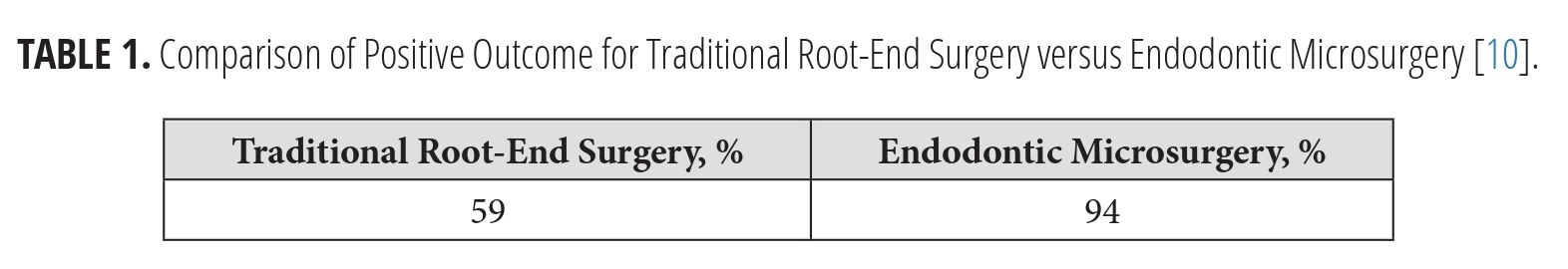
TABLE 1. Comparison of Positive Outcome for Traditional Root-End Surgery versus Endodontic Microsurgery [10].
Summarizing the research, it is possible to note that EM is 35% more successful procedure comparing to TRES [10].
Looking at these numbers, all conclusions are obvious. The future lies in the shift of many specialists involved in traditional root-end surgery to self-perform EM or referral to colleagues specializing in this microsurgical direction of dentistry. Having 9 years of experience in dentistry plus 19 years in oral and maxillofacial surgery, I finally want to say to my colleagues that no matter how many years we perform traditional surgical techniques like TRES, we always must rethink what is best for the patient. In sum, it is a pleasure to see how periapical surgery is evolving right in front of our eyes.
Oleksandr A. Nozhenko
Practice Limited to Oral and
Maxillofacial Surgery
Kyiv Regional Clinical Hospital
Kyiv, Ukraine
E-mail: alexdent03@gmail.com
REFERENCES (10)
-
Smith CS. “Alveolar abscess.” Am J Dent Sci. 1871;5(3rd series):289-300.
-
Gutmann JL, Gutmann MS. Historical perspectives on the evolution of surgical procedures in endodontics. J Hist Dent. 2010;58(1):1-42.
-
Baumann RR. How may the dentist benefit from the operating microscope? Quintessence Intern. 1977;5:17-18.
-
Apotheker H, Jako GJ. A microscope for use in dentistry. J Microsurg. 1981;3(1):7-10. https://doi.org/10.1002/micr.1920030104
-
Kim JE, Shim JS, Shin Y. A new minimally invasive guided endodontic microsurgery by cone beam computed tomography and 3-dimensional printing technology. Restor Dent Endod. 2019;44(3):e29. https://doi.org/10.5395%2Frde.2019.44.e29
-
Iandolo A, Abdellatif D, Barbosa AFA, et al. Confocal laser scanning microscopy evaluation of roots subjected to activation protocol in endodontic microsurgery. Aust Endod J. 2022;48(1):77-81. https://doi.org/10.1111/aej.12598
-
Tkachenko O, Volokitin A. A clinical case of endodontic microsurgery with a histological diagnosis of an apical scar. J Endod Microsurg. 2023;2:2-23. https://doi.org/10.23999/jem.2023.2.2
-
Fernández-Grisales R, Rojas WJ, Berruecos-Orozco C. Piezoelectric endodontic microsurgery with modified cortical window technique: a case report. J Endod Microsurg. 2023;2:34-40. https://doi.org/10.23999/jem.2023.2.4
-
Fesenko II. The time has come: Journal of Endodontic Microsurgery: a first peer-reviewed open access publication focused on microsurgery in endodontics. J Endod Microsurg. 2022;1:1-4. https://doi.org/10.23999/jem.2022.1.1
-
Setzer FC, Shah SB, Kohli MR, Karabucak B, Kim S. Outcome of endodontic surgery: a meta-analysis of the literature--part 1: comparison of traditional root-end surgery and endodontic microsurgery. J Endod. 2010;36(11):1757-1765. https://doi.org/10.1016/j.joen.2010.08.007